Types of Muscles
Gross Anatomy · Foundations
Introduction
Introduction to Muscle Types
Muscle tissue is a highly specialized tissue characterized by its ability to contract and generate force, driving locomotion, internal organ function, and structural stability. The human body contains hundreds of muscles, which are classified into three structurally and functionally distinct categories: skeletal muscle, cardiac muscle, and smooth muscle. Understanding these classifications is fundamental to studying human anatomy, histology, and physiology.
Study
Histological and Structural Classifications
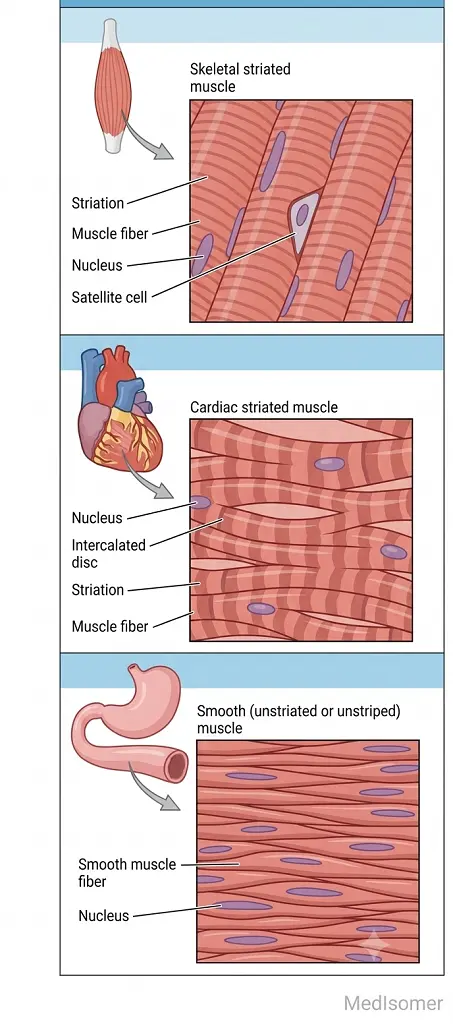
1. Skeletal Muscle: - Striated and voluntary. - Cells (muscle fibers) are long, cylindrical, and multinucleated, with nuclei peripherally located just beneath the sarcolemma. - The alternating dark (A bands) and light (I bands) striations are due to the highly organized arrangement of actin and myosin myofilaments into sarcomeres. 2. Cardiac Muscle: - Striated and involuntary. - Cells are short, branched, and typically contain a single, centrally located nucleus (occasionally binucleated). - Features unique junctional complexes known as intercalated discs, which contain gap junctions (for electrical coupling) and desmosomes (for mechanical anchorage). 3. Smooth Muscle: - Non-striated and involuntary. - Cells are spindle-shaped (fusiform) with a single, central nucleus. - Lacks organized sarcomeres; instead, actin and myosin filaments anchor to dense bodies, which act similarly to Z-discs, distributed throughout the cytoplasm and sarcolemma.
Functional and Functional Architecture
Skeletal muscles serve to move the skeleton, maintain posture, and stabilize joints under the direct control of the somatic nervous system. Cardiac muscle forms the myocardium of the heart walls, automatically and rhythmically pumping blood throughout the circulatory system via autonomic nervous regulation. Smooth muscle lines the walls of hollow visceral structures (such as blood vessels, the gastrointestinal tract, respiratory airways, and the bladder) to regulate luminal diameter and propagate substances via peristalsis under autonomic and endocrine influence.
Connective Tissue Coverings of Skeletal Muscle
Skeletal muscle organs are protected and compartmentalized by dense and loose connective tissue layers: - Epimysium: An outer layer of dense irregular connective tissue that surrounds the entire muscle belly. - Perimysium: A central connective tissue layer that groups individual muscle fibers into bundles called fascicles. - Endomysium: A delicate layer of reticular connective tissue that invests each individual muscle fiber, carrying capillaries and nerve axons.
Summary
Summary of Muscle Types
The three types of muscle cells vary based on voluntariness, structural striations, cell shape, and nuclei numbers. Skeletal muscles are voluntary, multinucleated, striated cylinders driving skeletal movement. Cardiac muscle is an involuntary, single-nucleated, branched structure containing intercalated discs dedicated to cardiac contraction. Smooth muscle consists of involuntary, single-nucleated, non-striated fusiform cells managing visceral operations. Structurally, skeletal muscle organization is sustained by three distinct layers of connective tissue: epimysium, perimysium, and endomysium.