Surface Anatomy
Gross Anatomy · Foundations
Introduction
Introduction to Surface Anatomy
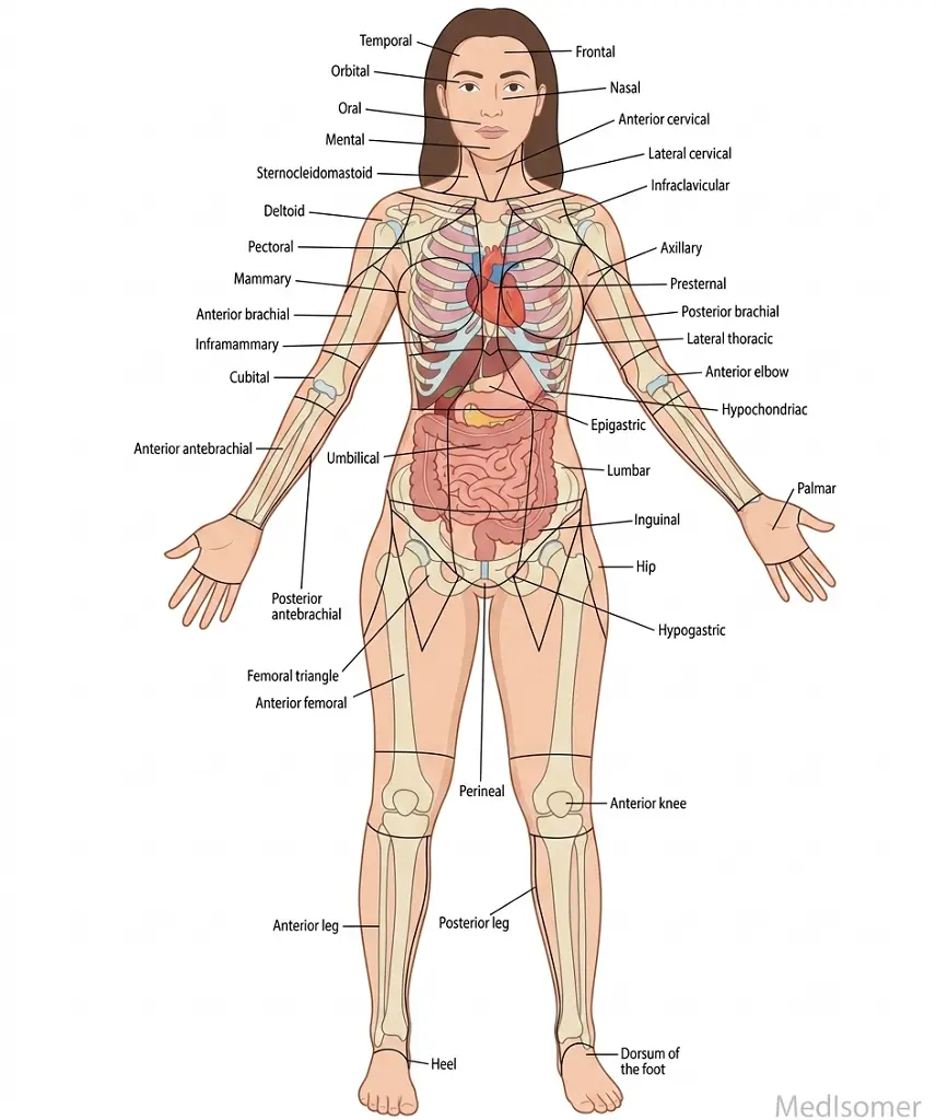
Surface anatomy (or superficial anatomy) is the study of the external features of the body which serve as superficial landmarks to locate deeper, internal structures. It forms the foundational bridge between gross anatomy and physical examination in clinical practice. By utilizing visual inspection and digital palpation, clinicians can accurately map underlying bones, muscles, nerves, and blood vessels without invasive procedures. Mastery of surface anatomy is indispensable for tasks ranging from finding the correct site for an intravenous line to performing emergency procedures like a cricothyroidotomy.
Study
Key Landmarks of the Head and Neck
In the head and neck, several prominent structures guide diagnostic and therapeutic interventions. The external occipital protuberance and zygomatic arch are key cranial reference points. In the midline of the neck, the thyroid cartilage ('Adam's apple') sits at the level of the C4-C5 vertebrae, serving as an anchor point. Immediately inferior to it lies the cricoid cartilage at the level of C6, marking the junction between the larynx and trachea, as well as the pharynx and esophagus. The cricothyroid membrane, located between these two cartilages, is the crucial site for emergency surgical airways.
Thoracic Landmarks and the Lines of Orientation
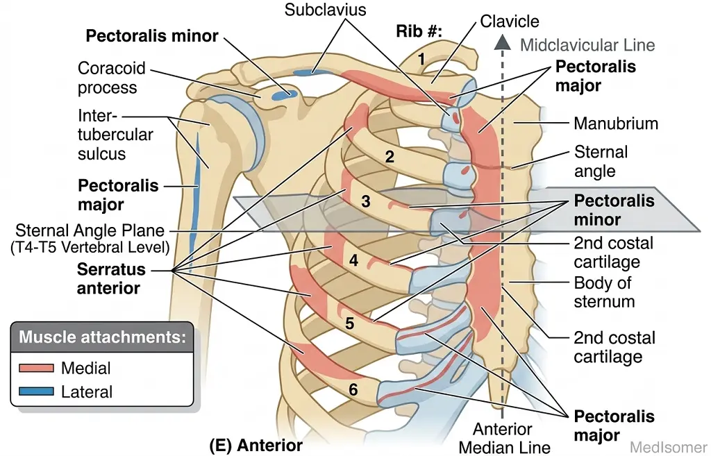
The thoracic wall relies on stable bony landmarks. The jugular notch (suprasternal notch) sits at the superior border of the manubrium. Moving inferiorly, the manubriosternal joint forms a distinct ridge known as the Sternal Angle (Angle of Louis). This clinical milestone marks the level of the 2nd costal cartilages, the T4-T5 intervertebral disc space, the bifurcation of the trachea, and the transition of the aortic arch. Vertical reference lines are projected across these landmarks to isolate thoracic zones: the midclavicular line (dropping vertically from the midpoint of the clavicle), the anterior axillary line (along the anterior axillary fold), and the midaxillary line (dropping from the apex of the axilla).
Abdominal Topography and Quadrants
The abdomen is superficially divided into mapping zones to localize pain and pathology. The simplest method uses two perpendicular planes intersecting at the umbilicus (L3-L4 level) to create four quadrants: Right Upper (RUQ), Left Upper (LUQ), Right Lower (RLQ), and Left Lower (LLQ). Alternatively, a nine-region system uses two vertical midclavicular lines and two horizontal planes: the subcostal plane (inferior margin of 10th costal cartilage, L3 level) and the transtubercular plane (iliac tubercles, L5 level). Critical landmarks within these regions include McBurney's point—located one-third of the distance from the right anterior superior iliac spine (ASIS) to the umbilicus—which signifies the base of the appendix, and the transpyloric plane of Addison (midway between the jugular notch and pubic symphysis at L1), which cuts through the pylorus, hila of kidneys, and fundus of the gallbladder.
Extremity Landmarks for Clinical Access
In the upper limb, the cubital fossa is an anterior triangular depression bounded by the pronator teres and brachioradialis muscles; it contains the median nerve, brachial artery, and biceps tendon, while the superficial median cubital vein lies overhead, serving as the primary site for venipuncture. On the posterior wrist, extending the thumb reveals the anatomical snuffbox, bounded medially by the extensor pollicis longus and laterally by the abductor pollicis longus and extensor pollicis brevis. The scaphoid bone forms its floor, and the radial artery runs through it. In the lower limb, the femoral triangle in the upper anterior thigh is bounded by the inguinal ligament, sartorius, and adductor longus. It contains the Femoral Nerve, Artery, and Vein (ordered from lateral to medial: NAVEL), accessible just inferior to the mid-inguinal point.
Summary
Summary of High-Yield Clinical Correlates
Surface anatomy systematically links superficial visual boundaries to underlying clinical anatomy. Remember these vital relationships: • Sternal Angle: 2nd rib, T4-T5 level, separates superior and inferior mediastinum. • Cricothyroid Membrane: Inframandibular neck midline, site for emergency cricothyroidotomy. • McBurney's Point: 1/3 from right ASIS to umbilicus, localizes maximum tenderness in acute appendicitis. • Anatomical Snuffbox: Floor contains the scaphoid bone; site of palpatory tenderness in scaphoid fractures. • Femoral Triangle: Houses the femoral pulse directly midpoint between the ASIS and pubic symphysis.