Scapula
Gross Anatomy · Osteology
Introduction
Introduction to the Scapula
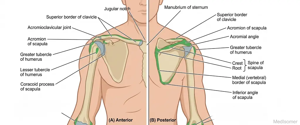
The scapula, commonly referred to as the shoulder blade, is a large, flat, triangular bone positioned on the posterolateral aspect of the thoracic cage. Extending from the second to the seventh ribs, it serves as a crucial anchor for the upper limb, connecting the appendicular skeleton to the axial skeleton via functional muscular attachments and its articulation with the clavicle. As a key component of the pectoral girdle, the scapula provides structural stability while affording the upper limb an extraordinary range of motion.
Study
Anatomical Landmarks and Borders
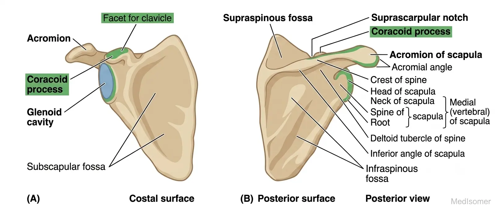
The scapula features two surfaces (costal and posterior), three borders (superior, medial, and lateral), and three angles (superior, inferior, and lateral). 1. Surfaces: - Costal (Anterior) Surface: This surface faces the ribs and presents a large, concave depression known as the subscapular fossa, which provides attachment for the subscapularis muscle. - Posterior Surface: This surface is uneven and divided into two sections by a prominent, transverse projection called the spine of the scapula. The smaller region above the spine is the supraspinous fossa, while the larger region below it is the infraspinous fossa. These fossae house the supraspinatus and infraspinatus muscles, respectively. The spine terminates laterally as a flattened, expanded process known as the acromion, which projects anterolaterally and articulates with the clavicle. 2. Borders and Angles: - Superior Border: The shortest and thinnest border, featuring the suprascapular notch near the base of the coracoid process. This notch is converted into a foramen by the superior transverse scapular ligament, transmitting the suprascapular nerve. - Medial (Vertebral) Border: A thin border running parallel to the vertebral column, serving as an insertion site for muscles like the rhomboids and levator scapulae. - Lateral (Axillary) Border: The thickest border, running from the inferior angle up to the glenoid cavity. It contains the infraglenoid tubercle at its upper end. - Lateral Angle and Glenoid Cavity: The lateral angle is truncated and expanded to form the glenoid cavity. This shallow, pyriform (pear-shaped) articular surface articulates with the head of the humerus to form the glenohumeral (shoulder) joint. Immediately superior to the glenoid cavity is the supraglenoid tubercle, and inferior to it is the infraglenoid tubercle. - Coracoid Process: A thick, hook-like bony projection extending anterolaterally from the superior border just medial to the glenoid cavity. It serves as an essential attachment site for ligaments and muscles.
Muscular Attachments and Clinical Correlations
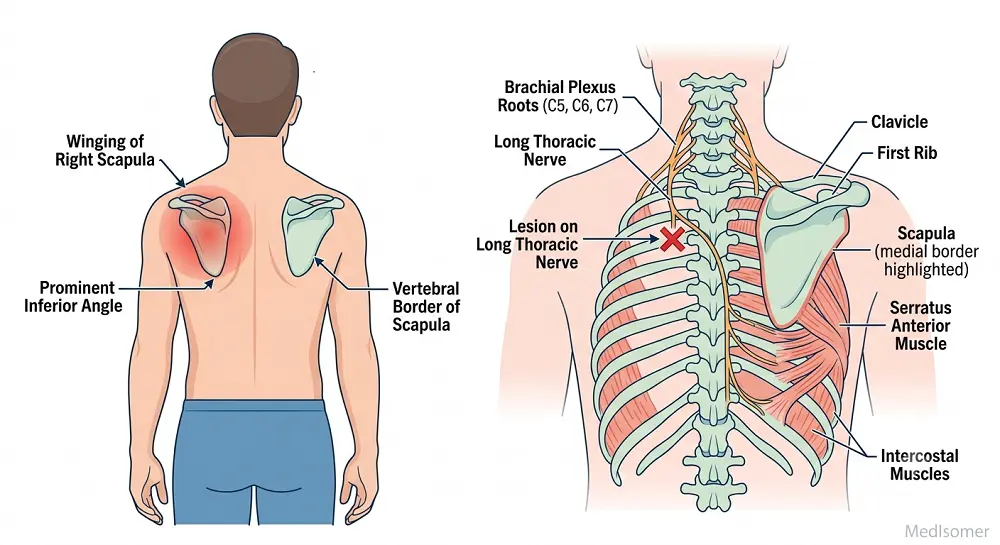
The scapula serves as the origin or insertion point for 17 muscles, making it highly dynamic. Key Muscular Attachments: - Rotator Cuff Muscles: Four muscles maintain glenohumeral stability. The Supraspinatus originates from the supraspinous fossa, Infraspinatus from the infraspinous fossa, Teres Minor from the upper lateral border, and Subscapularis from the subscapular fossa. - Serratus Anterior: Inserts onto the anterior aspect of the medial border. It tethers the scapula against the thoracic wall and protracts it during punching or reaching movements. - Trapezius and Deltoid: The trapezius inserts along the superior border of the spine and acromion, while the deltoid takes origin from the inferior border of the spine and acromion. - Biceps Brachii: The long head originates from the supraglenoid tubercle, and the short head originates from the tip of the coracoid process. - Triceps Brachii: The long head originates from the infraglenoid tubercle. Clinical Insights: - Winging of the Scapula: Injury to the long thoracic nerve (which paralyzes the serratus anterior muscle) causes the medial border of the scapula to project posteriorly like a wing when the patient pushes against a wall. - Scapular Fractures: Due to protection provided by surrounding thick muscle beds, fractures of the scapula are relatively rare and usually result from high-energy severe trauma (e.g., motor vehicle accidents), often associated with rib fractures or lung injuries.
Summary
Topic Summary
The scapula is a flat, triangular bone forming the posterior segment of the pectoral girdle. Its critical features include the costal surface (subscapular fossa) and the posterior surface, which is split by the scapular spine into the supraspinous and infraspinous fossae. The spine culminates laterally into the acromion, which articulates with the clavicle at the acromioclavicular joint. The lateral angle houses the shallow glenoid cavity for articulation with the humeral head, flanked by the coracoid process anterosuperiorly. Acting as a platform for 17 muscles, including the rotator cuff group and the serratus anterior, structural or neural disruptions can lead to functional clinical deficits such as scapular winging.