Principles of Kinesiology
Gross Anatomy · Foundations
Introduction
Introduction to Kinesiology
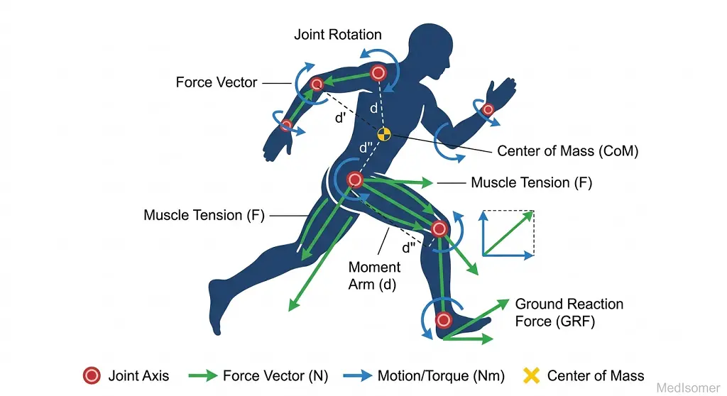
Kinesiology is the scientific study of human body movement. It integrates principles from anatomy, physiology, and biomechanics to analyze how the musculoskeletal system interacts to produce coordinated, efficient motion. For medical and anatomical studies, understanding kinesiology is essential for assessing joint mechanics, muscle function, gait abnormalities, and rehabilitative pathways.
Study
Biomechanical Levers and Mechanical Advantage
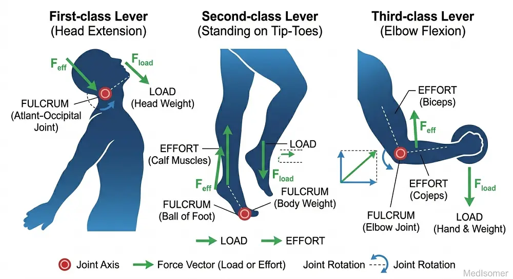
The musculoskeletal system functions primarily as a system of levers. A lever consists of a rigid bar (bone), a fulcrum (joint), an effort or force (muscle contraction), and a load or resistance (weight of the limb or external object). Levers are classified into three types based on the relative positions of these components: 1. First-Class Levers: The fulcrum is positioned between the effort and the load (E-F-L). An anatomical example is the atlanto-occipital joint during head extension, where the joint is the fulcrum, the posterior neck muscles provide the effort, and the facial weight is the load. 2. Second-Class Levers: The load is positioned between the fulcrum and the effort (F-L-E). This setup provides a mechanical advantage greater than 1, meaning less effort is required to move a heavy load. An anatomical example is plantarflexion at the metatarsophalangeal joints when standing on tip-toes; the toes act as the fulcrum, the body weight via the tibia is the load, and the gastrocnemius/soleus complex provides the effort. 3. Third-Class Levers: The effort is positioned between the fulcrum and the load (F-E-L). This is the most common lever system in the human body. While it operates at a mechanical disadvantage (requiring more muscle force than the resistance being moved), it maximizes speed and range of motion. An anatomical example is elbow flexion by the biceps brachii, where the elbow joint is the fulcrum, the biceps insertion on the radial tuberosity is the effort, and the forearm/hand weight is the load.
Kinematic Chains and Joint Kinematics
Movement occurs through kinematic chains, which describe a series of overlapping segments connected via joints. These are split into two categories: * Open Kinematic Chain (OKC): The distal segment of the limb is free to move in space while the proximal segment remains fixed. Examples include knee extension on a leg machine or throwing a ball. OKC movements allow isolated muscle activation and high-velocity movements. * Closed Kinematic Chain (CKC): The distal segment of the limb is fixed or anchored to an immovable surface (like the floor). Consequently, movement at one joint automatically induces predictable movements at adjacent joints. Examples include squats, push-ups, or the stance phase of gait. CKC exercises recruit multiple joint complexes and increase joint stability through axial loading. Joint motion itself is classified into Osteokinematics (the visible gross movement of bones in space, such as flexion or abduction) and Arthrokinematics (the microscopic, involuntary movements occurring between joint articulating surfaces, categorized as rolling, sliding/gliding, and spinning).
Summary
Summary of Core Concepts
Principles of kinesiology combine mechanical vectors with structural anatomy. Human movement relies heavily on third-class lever configurations, trading mechanical force efficiency for maximized displacement speed and range of motion. Functional movements are coordinated via open and closed kinematic chains, supported at the joint interface by a fine balance between gross osteokinematic pathways and subtle arthrokinematic glides, rolls, and spins.