Planes of the Body
Gross Anatomy · Foundations
Introduction
Introduction to Anatomical Planes
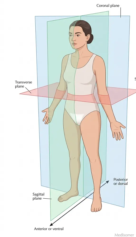
In medical and anatomical sciences, describing the relative positions of structures, movements, or imaging slices requires a standardized reference framework. This framework begins with the standard anatomical position: an individual standing erect, facing forward, upper limbs at the sides, palms facing forward, and feet flat on the floor pointing ahead. To describe internal geography, three-dimensional spaces are sliced by imaginary flat surfaces known as anatomical planes. Understanding these planes is foundational for clinical examinations, surgical approaches, and interpreting radiological imaging like CT, MRI, and ultrasound.
Study
The Three Cardinal Anatomical Planes
1. Sagittal (Median) Plane: A vertical plane running from anterior (front) to posterior (back). It divides the body or an organ into right and left portions. The specific sagittal plane that lies exactly in the midline and splits the body into equal right and left halves is called the midsagittal or median plane. Parasagittal planes run parallel to the median plane, dividing the body into unequal right and left portions. 2. Frontal (Coronal) Plane: A vertical plane oriented perpendicular to the sagittal plane. It divides the body or an organ into anterior (ventral/front) and posterior (dorsal/back) portions. For example, a coronal cut through the head would separate the face from the back of the skull. 3. Transverse (Horizontal / Axial) Plane: A horizontal plane oriented perpendicular to both the sagittal and coronal planes. It divides the body or an organ into superior (upper) and inferior (lower) portions. In clinical medicine, this is commonly referred to as an axial plane, which is the standard orientation for presenting traditional CT slices.
Oblique Planes and Cross-Sectional Geometry
Any plane that passes through the body or an organ at an angle other than a 90-degree angle to the horizontal or vertical axes is classified as an oblique plane. These are common when imaging structures that do not lie cleanly along standard cardinal axes, such as the heart (long and short axes) or the appendix. When an organ is sliced, the cut surfaces are called sections. A longitudinal section is a cut along the long axis of a structure (which can be sagittal or coronal), whereas a cross-section is a cut perpendicular to the long axis of a structure.
Clinical and Radiographic Relevance
Modern imaging modalities rely strictly on these planes for diagnostic clarity. Computed Tomography (CT) scans natively capture data in the axial (transverse) plane but are routinely reconstructed into coronal and sagittal views. Magnetic Resonance Imaging (MRI) can natively acquire images in any orthogonal or oblique plane. Ultrasonography depends heavily on the orientation of the transducer probe, requiring clinicians to actively manipulate the probe along sagittal, transverse, or oblique planes to trace dynamic anatomical structures like blood vessels, nerve bundles, or organs.
Summary
Summary of Core Concepts
Anatomical planes are essential geometric references used to describe body sections and radiological imaging. The sagittal plane divides the body into right and left sides (midsagittal for equal halves); the coronal or frontal plane divides it into anterior and posterior parts; and the transverse or axial plane cuts horizontally to yield superior and inferior sections. Oblique planes slice at non-right angles. Mastery of these concepts is indispensable for accurately interpreting diagnostic imaging (CT, MRI, Ultrasound) and executing surgical dissections.