Osteology: General Principles
Gross Anatomy · Foundations
Introduction
Introduction to Osteology and the Skeletal System
Osteology is the scientific study of bones, which serve as the structural framework of the human body. Far from being static, inert structures, bones are dynamic, living tissues that continuously remodel themselves in response to mechanical stress and metabolic demands. Together with cartilage, they form the skeletal system, which serves indispensable functions: providing mechanical support, facilitating locomotion by acting as levers for muscles, shielding vital internal organs from injury, housing hematopoietic tissue for blood cell production, and serving as a metabolic reservoir for vital minerals such as calcium and phosphorus.
Study
Functions of the Skeletal System
The skeletal system possesses five primary functions. 1. Support: It provides a rigid framework that supports the body's weight and maintains its shape against gravity. 2. Protection: Bony enclosures shield vulnerable internal organs; for example, the cranium protects the brain, the vertebral column protects the spinal cord, and the thoracic cage protects the heart and lungs. 3. Movement: Bones serve as lever arms, and joints serve as fulcrums. Skeletal muscles attach to bones via tendons, and muscle contraction creates torque that moves the skeleton. 4. Mineral Homeostasis: Bones store roughly 99% of the body's calcium and 85% of its phosphorus. They can dynamically release or store these ions to maintain tight serum levels under endocrine control. 5. Hematopoiesis: The red bone marrow located within the trabecular spaces of specific bones is the primary site of blood cell production (erythrocytes, leukocytes, and platelets).
Structural Divisions: Axial vs. Appendicular Skeleton
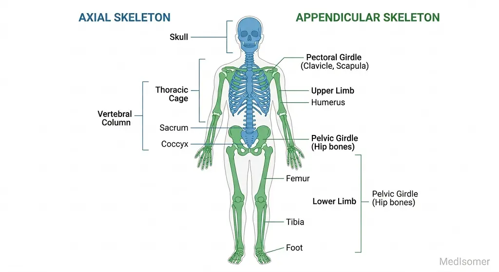
The adult human skeleton typically consists of 206 distinct bones, which are organized into two major structural divisions: the Axial Skeleton and the Appendicular Skeleton. The axial skeleton forms the central core axis of the body and comprises 80 bones: the skull (cranium and facial bones), the vertebral column (cervical, thoracic, lumbar, sacral, and coccygeal vertebrae), the hyoid bone, the auditory ossicles, and the thoracic cage (ribs and sternum). The appendicular skeleton consists of 126 bones associated with the upper and lower limbs and their attachments to the trunk: the pectoral girdles (scapulae and clavicles), the pelvic girdle (hip bones), and the bones of the arms, forearms, wrists, hands, thighs, legs, ankles, and feet.
Gross Anatomical Classification of Bones
Bones are classified into five distinct categories based on their gross shape and configuration: • Long Bones: Characterized by a length that exceeds their width, possessing a central shaft (diaphysis) and two expanding ends (epiphyses). Examples include the femur, humerus, radius, ulna, tibia, fibula, metacarpals, metatarsals, and phalanges. • Short Bones: Roughly cuboidal in shape, having nearly equal length, width, and thickness. They consist of a core of spongy bone surrounded by a thin layer of compact bone. Examples include the carpal bones of the wrist and tarsal bones of the ankle. • Flat Bones: Thin, parallel-layered structures composed of two plates of compact bone enclosing a central layer of spongy bone (known as the diploë in cranial bones). They provide extensive surface area for muscle attachment and protection. Examples include the parietal and frontal bones of the skull, the sternum, the ribs, and the scapulae. • Irregular Bones: Possess complex, unclassifiable shapes that do not fit into other categories. Examples include the vertebrae, the sacrum, the ethmoid, and the sphenoid bones. • Sesamoid Bones: Specialized bones embedded within tendons where significant friction, tension, and physical stress occur. They protect tendons from excessive wear and tear and mechanically alter the angle of muscle pull to improve leverage. The patella (kneecap) is the largest and most constant sesamoid bone in the body; others occur frequently in the tendons of the hand and foot.
Anatomy of a Typical Long Bone
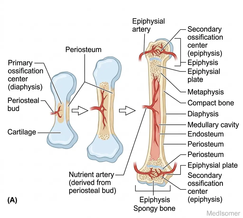
A classic long bone provides an ideal template to study gross bone structure. It consists of the following key anatomical regions: • Diaphysis: The elongated, cylindrical shaft of the bone. It is composed primarily of dense, heavy compact bone surrounding a central hollow chamber. • Medullary (Marrow) Cavity: The space within the diaphysis. In adults, it is filled with yellow bone marrow, which consists primarily of adipose tissue; in infants, it contains hematopoietically active red bone marrow. • Epiphyses: The proximal and distal extremities of the bone. They are typically bulbous expansions composed of an outer shell of compact bone filling an internal framework of spongy (trabecular) bone. The spaces within the spongy bone of certain adult epiphyses contain red bone marrow. • Metaphysis: The transitional region connecting the diaphysis to the epiphysis. In a growing bone, this region contains the epiphyseal plate (growth plate), a layer of hyaline cartilage where interstitial longitudinal bone growth occurs. When growth ceases, this cartilage ossifies entirely, leaving behind a bony remnant called the epiphyseal line. • Articular Cartilage: A thin layer of hyaline cartilage covering the joint surfaces of the epiphyses. It minimizes friction and absorbs mechanical shocks within synovial joints. It lacks a perichondrium and has limited regenerative capacity. • Periosteum: A tough, double-layered fibrous connective tissue sheath that covers the external surface of the bone wherever it is not covered by articular cartilage. It consists of an outer fibrous layer (dense irregular connective tissue providing attachment for tendons and ligaments) and an inner osteogenic layer (containing osteoprogenitor cells, osteoblasts, and osteoclasts critical for bone growth, remodeling, and repair). The periosteum is anchored to the underlying bone by thick bundles of collagen fibers called perforating (Sharpey's) fibers. It is richly supplied with nociceptive nerve fibers, making it highly sensitive to trauma. • Endosteum: A thin, vascular membrane that lines the internal medullary cavity, the osteon canals, and covers the trabeculae of spongy bone. It is a single cell layer thick and contains osteoprogenitor cells, osteoblasts, and osteoclasts, playing an essential role in bone growth and remodeling.
Microscopic Organization of Bone Tissue
Bone tissue (osseous tissue) is a specialized connective tissue characterized by a mineralized extracellular matrix. It exists in two microscopic structural formats: compact bone and spongy bone. Compact (Cortical) Bone forms the dense, protective outer cortex of all bones. Its structural and functional unit is the Osteon, or Haversian System. Each osteon is an elongated cylinder aligned parallel to the long axis of the bone, acting like a weight-bearing pillar. An osteon consists of: • Central (Haversian) Canal: Runs longitudinally through the center of the osteon, containing blood vessels, lymphatic vessels, and nerve fibers. • Concentric Lamellae: Rings of calcified extracellular matrix arranged concentrically around the central canal. • Lacunae: Tiny spaces situated between adjacent lamellae, each housing a mature bone cell called an osteocyte. • Canaliculi: Microscopic, radiating tunnels that link adjacent lacunae. This network allows for the rapid transport of nutrients throughout the dense, impermeable mineralized matrix. • Perforating (Volkmann's) Canals: Transverse or oblique channels that run perpendicular to the long axis of the bone. They carry blood vessels and nerves from the periosteum into the deeper bone tissue. • Interstitial Lamellae: Remnants of older, partially destroyed osteons that fill the spaces between intact, functional osteons. • Circumferential Lamellae: Large rings of bone matrix that extend around the entire inner and outer circumferences of the diaphysis. Spongy (Trabecular or Cancellous) Bone does not contain true osteons. Instead, it consists of an open, irregular lattice of branching bony spicules termed trabeculae. The trabeculae are precisely aligned along lines of mechanical stress to distribute weight efficiently.
The Bone Matrix and Bone Cells
The extracellular matrix of bone tissue consists of an organic component and an inorganic component. The organic matrix (osteoid) constitutes about one-third of the bone mass and is secreted by osteoblasts. It is composed predominantly of type I collagen fibers. Collagen gives bone its high tensile strength and flexibility. The inorganic matrix makes up the remaining two-thirds and consists of mineral salts, primarily hydroxyapatite. This component imparts exceptional hardness and compressive strength. Four distinct cell types maintain bone tissue: 1. Osteoprogenitor Cells: Mitotically active stem cells found in the periosteum and endosteum. 2. Osteoblasts: Bone-building cells that synthesize and secrete the organic components of the matrix. 3. Osteocytes: Mature bone cells trapped in lacunae that act as mechanosensors. 4. Osteoclasts: Massive, multinucleated cells responsible for bone resorption.
Bone Development and Growth (Ossification)
Osteogenesis, or ossification, is the process of bone formation. All bones derive initially from embryonic mesenchyme via two distinct pathways: • Intramembranous Ossification: Bone develops directly within mesenchymal connective tissue without a cartilaginous intermediate. This forms flat bones of the skull and the clavicle. • Endochondral Ossification: Bone replaces an existing model of hyaline cartilage. This process forms most bones of the skeleton. Bones grow via two concurrent processes: • Longitudinal (Interstitial) Growth: Occurs at the epiphyseal plates of long bones, driving lengthening. • Appositional Growth: Increases the width or thickness of bones as osteoblasts in the periosteum secrete new matrix while osteoclasts in the endosteum resorb bone.
Bone Remodeling and Calcium Homeostasis
Bone remodeling is a lifelong process preservation structural integrity and maintaining calcium balance. It is governed by Wolff's Law (mechanical stress) and hormonal loops. When blood calcium levels drop, Parathyroid Hormone (PTH) is released, stimulating bone resorption to release calcium. Conversely, when blood calcium levels rise, Calcitonin is secreted, inhibiting osteoclast activity to shift excess calcium back into the bony matrix.
Summary
Summary of Key Osteology Principles
• Osteology encompasses the structural, mechanical, and metabolic properties of bone tissue. • The skeletal system is divided into axial and appendicular skeletons. • Bones are categorized by shape into long, short, flat, irregular, and sesamoid classes. • Long bones possess a distinct diaphysis, epiphyses, and medullary cavity. • Compact bone is organized into osteons, while spongy bone forms trabeculae. • The matrix contains organic collagen (flexibility) and inorganic hydroxyapatite (hardness). • Bone is maintained by osteoprogenitor cells, osteoblasts, osteocytes, and osteoclasts. • Development occurs via intramembranous or endochondral ossification. • Systemic calcium balance is regulated by PTH and Calcitonin.