Myology: General Principles
Gross Anatomy · Foundations
Introduction
Introduction to Myology and Muscle Architecture
Myology is the branch of anatomy dedicated to the scientific study of muscles, their structural organization, attachments, and functional mechanics. Muscles are highly specialized biological engines that convert chemical energy, stored in the form of adenosine triphosphate (ATP), into mechanical work and movement. Within the human body, the muscular system serves as the primary driver of locomotion, posture maintenance, internal material transport (such as blood propulsion and peristalsis), and thermogenesis.
Understanding the general principles of myology requires a comprehensive view of how individual muscle cells, or fibers, are bundled and integrated with connective tissue frameworks. This architectural design not only ensures structural integrity during high-stress contractions but also determines the excursion range and absolute power output of each distinct muscle group. This module explores the core histological variations, structural layers, dynamic mechanisms of contraction, and structural adaptations that define human musculature.
Study
1. Histological Classification of Muscle Tissue
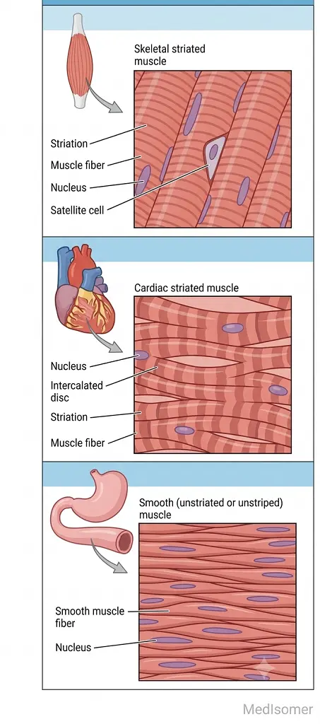
The human muscular system is categorized into three distinct histological and functional types: skeletal, cardiac, and smooth muscle. Skeletal muscle is striated, multinucleated, and primarily under voluntary control by the somatic nervous system. Its cells are elongated cylinders formed by the fusion of embryonic myoblasts. Cardiac muscle, found exclusively in the myocardium, is also striated but operates involuntarily under autonomic and intrinsic pacemaking control. Structurally, cardiac myocytes are branched, possess one or two centrally located nuclei, and are linked end-to-end by specialized junctional complexes known as intercalated discs, which contain gap junctions for rapid electrical coupling. Smooth muscle is non-striated, mononucleated, spindle-shaped, and located within the walls of hollow visceral organs and blood vessels. It operates involuntarily, exhibiting slow, sustained contractions modulated by the autonomic nervous system, hormones, and local metabolic factors.
2. Connective Tissue Framework of Skeletal Muscle
Skeletal muscles are not merely collections of isolated fibers; they are meticulously organized by hierarchical layers of dense and loose connective tissue. The entire muscle belly is invested by an outer sheath of dense irregular connective tissue called the epimysium, which separates the muscle from surrounding tissues and reduces friction. Deep to the epimysium, the muscle is divided into internal bundles of fibers called fascicles. Each fascicle is wrapped by a collagenous connective tissue layer known as the perimysium, which houses major blood vessels and nerves supplying the fibers. Within each fascicle, individual muscle fibers are enveloped by a delicate layer of reticular fibers and loose connective tissue called the endomysium. The endomysium contains a rich capillary network and fine nerve terminals. At the ends of the muscle belly, these three continuous connective tissue sheaths merge to form deep fascia, tendons, or broad aponeuroses that anchor the muscle to bone or soft tissue.
3. Microscopic Structure and the Sarcomere
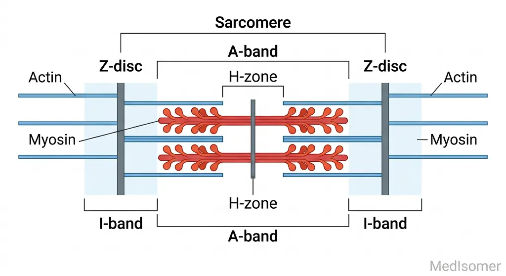
An individual skeletal muscle fiber contains thousands of longitudinal, cylindrical structures called myofibrils. Myofibrils exhibit a highly organized pattern of alternating light and dark bands, reflecting the arrangement of microscopic myofilaments. The basic functional and contractile unit of a myofibril is the sarcomere, delimited longitudinally by two successive Z-discs (Z-lines). Thin filaments, composed primarily of actin, tropomyosin, and the troponin complex, are anchored directly to the Z-discs. Thick filaments, consisting of highly organized myosin molecules with projecting globular heads, occupy the center of the sarcomere. The dark band is designated as the A-band and corresponds to the full length of the thick myosin filaments, including zones of overlap with thin filaments. The light band is the I-band, which contains only thin actin filaments and spans across the Z-disc into adjacent sarcomeres. Within the center of the A-band lies the H-zone, a lighter region containing only thick filaments. In the exact center of the H-zone is the M-line, formed by structural proteins that anchor and stabilize the thick filaments.
4. The Sliding Filament Mechanism and Excitation-Contraction Coupling
Muscle contraction occurs via the sliding filament mechanism. Upon stimulation, myosin heads bind to active sites on actin filaments, forming cross-bridges. Utilizing energy derived from ATP hydrolysis, the myosin heads undergo a conformational change known as the power stroke, pulling the thin actin filaments inward toward the M-line. During this process, the individual myofilaments do not shorten; instead, the sarcomere shortens as the thin filaments slide past the thick filaments, causing the H-zone and I-bands to narrow or disappear completely, while the width of the A-band remains strictly constant.
This mechanical event is initiated by excitation-contraction coupling. An action potential arriving at the neuromuscular junction triggers the release of acetylcholine (ACh) into the synaptic cleft. ACh binds to nicotinic receptors on the motor endplate, generating a localized depolarization that propagates across the sarcolemma (muscle cell membrane) and deep into the interior of the fiber via transverse tubules (T-tubules). The T-tubules run adjacent to the terminal cisternae of the sarcoplasmic reticulum (SR)—a specialized intracellular calcium storage network. A T-tubule and its two flanking terminal cisternae form a structural triad. The depolarization activates voltage-gated L-type calcium channels (dihydropyridine receptors, DHPR) in the T-tubule membrane, which mechanically open calcium-release channels (ryanodine receptors, RyR) in the SR membrane. Calcium ions flood the sarcoplasm and bind to troponin C on the thin filaments. This binding induces a conformational shift in tropomyosin, moving it away from the active sites on actin, allowing myosin heads to bind and initiate the cross-bridge cycle.
5. Fascicular Architecture and Functional Mechanics
The orientation and arrangement of muscle fascicles relative to the central tendon profoundly influence a muscle's physiological properties. Parallel muscles have fascicles that run parallel to the long axis of the muscle (e.g., rectus abdominis, sartorius). This layout permits a maximal range of motion and a high velocity of contraction but generates less absolute force. Pennate muscles have fascicles that attach obliquely to their central tendon, resembling a feather. They can be unipennate (extensor digitorum longus), bipennate (rectus femoris), or multipennate (deltoid). Because pennate muscles pack a significantly larger number of muscle fibers into a given volume, they possess a much higher physiological cross-sectional area (PCSA), allowing them to generate immense force, though they suffer from a restricted range of movement and lower shortening speed.
Summary
Summary of Core Principles
Myology integrates macroscopic architectural design with microscopic molecular mechanics. Skeletal muscle relies on a continuous connective tissue framework (epimysium, perimysium, and endomysium) to harness the microscopic shortening of its sarcomeres and transmit it as macroscopic pull via tendons. Contraction is driven by calcium release at the triad, enabling the sliding filament mechanism where actin slides past myosin without changing filament length. Ultimately, the mechanical capabilities of a muscle are defined by its fascicular geometry: parallel configurations favor range of motion and speed, whereas pennate configurations optimize physiological cross-sectional area to maximize absolute force production.