Humerus
Gross Anatomy · Osteology
Introduction
Introduction to the Humerus
The humerus is the longest and largest bone of the upper limb, forming the structural framework of the arm (brachium). It articulates proximally with the scapula at the glenohumeral joint and distally with the radius and ulna at the elbow joint. Serving as a crucial mechanical lever, the humerus facilitates a vast range of upper extremity motions, allowing the hand to be dynamically positioned in space. Beyond its mechanical role, it features distinct anatomical regions susceptible to specific neurovascular injuries, making a comprehensive understanding of its features indispensable for both clinical examination and surgical management.
Study
Proximal Anatomy and Bony Landmarks
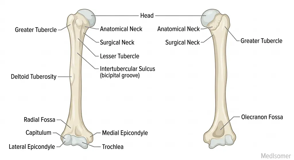
The proximal end of the humerus features the smooth, hemispherical head, which faces medially, superiorly, and posteriorly to articulate with the glenoid cavity of the scapula. Immediately bordering the articular margin is the anatomical neck. Distal to the head are two prominent muscular attachments: the larger, laterally placed greater tubercle and the smaller, anteriorly directed lesser tubercle. Separating these structures is the intertubercular sulcus (bicipital groove), which lodges the long head of the biceps brachii tendon and its synovial sheath. The transition zone where the expanded proximal end tapers into the cylindrical shaft is designated as the surgical neck, so named because it is a frequent site of fracture and lies in intimate contact with the axillary nerve and posterior circumflex humeral artery.
The Humeral Shaft
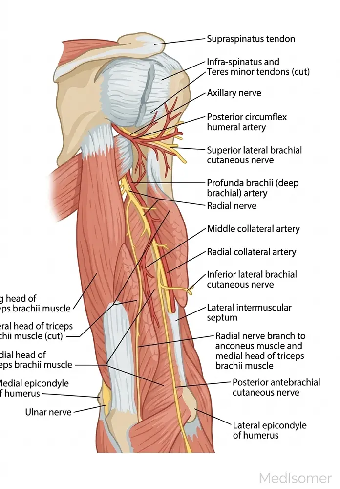
The shaft (diaphysis) of the humerus progressively changes from a cylindrical shape proximally to a triangular cross-section distally. On its lateral surface, near the midpoint, lies the V-shaped deltoid tuberosity, which serves as the insertion site for the deltoid muscle. Running obliquely along the posterior and lateral aspect of the shaft is the radial (spiral) groove. This groove transmits the radial nerve and the profunda brachii artery (deep artery of the arm). Fractures along the middle third of the humeral shaft can compress or lacerate the radial nerve within this groove, leading to a classic presentation of 'wrist drop' due to paralysis of the extensor muscles of the forearm.
Distal End and Articular Surfaces
The distal humerus is flattened anteroposteriorly and features non-articular and articular components. The non-articular landmarks include the prominent medial epicondyle and the smaller lateral epicondyle, both of which serve as major attachment sites for forearm muscles (the common flexor and common extensor origins, respectively). The ulnar nerve passes directly behind the medial epicondyle in the ulnar groove, making it highly vulnerable to compression or direct trauma. The articular surfaces comprise the lateral, pulley-like capitulum, which articulates with the head of the radius, and the medial, spool-shaped trochlea, which articulates with the trochlear notch of the ulna. Depressions accommodate forearm bones during movement: the anterior coronoid fossa (for the ulna during flexion), the radial fossa (for the radius during flexion), and the deep posterior olecranon fossa (which accommodates the olecranon process of the ulna during full elbow extension).
Clinical Correlations and Fractures
The humerus is highly clinically relevant due to predictable patterns of neurovascular injury associated with fractures at specific regions. 1. Proximal/Surgical Neck Fractures: Common in elderly patients with osteoporosis following a fall on an outstretched hand; carries a high risk of damage to the axillary nerve (resulting in deltoid atrophy and loss of sensation over the lateral arm) and the posterior circumflex humeral artery. 2. Mid-shaft Fractures: Frequently damage the radial nerve in the spiral groove, leading to wrist drop. 3. Supracondylar Fractures: Occur predominantly in children following hyperextension injuries of the elbow. The proximal bone fragment can be displaced anteriorly, injuring the median nerve and brachial artery, potentially leading to Volkmann's ischemic contracture.
Summary
Key Summary of Humeral Anatomy
The humerus bridges the shoulder and elbow, possessing a proximal head, a rigid shaft, and a specialized distal condyle. Proximally, the greater and lesser tubercles provide leverage for rotator cuff muscles, while the surgical neck represents a major structural vulnerability point for the axillary nerve. The shaft houses the deltoid tuberosity laterally and the radial groove posteriorly, which protects the radial nerve. Distally, the capitulum and trochlea form the articular core of the elbow joint, flanked by epicondyles that orchestrate forearm muscle dynamics. Mastery of humeral structures, coupled with their respective neurovascular relations (Axillary at the surgical neck, Radial at the mid-shaft, and Ulnar/Median at the distal end), forms the basis for managing upper limb pathology and trauma.