Arthrology: General Principles
Gross Anatomy · Foundations
Introduction
Introduction to Arthrology
Arthrology is the branch of anatomy dedicated to the scientific study of joints, or articulations. A joint is formed wherever two or more bones, or components of bone and cartilage, meet one another. Joints serve two contrasting yet vital evolutionary functions: they secure our skeletal components firmly together while simultaneously providing the structural flexibility required to facilitate synchronized movement. Whether it is the immobile fusion lines of the skull or the highly mobile architecture of the shoulder, understanding the principles of arthrology is foundational to mastering human biomechanics and clinical orthopedics.
Study
Classification of Joints
Joints are classified using two distinct but complementary systems: structural (based on anatomical features) and functional (based on the degree of movement permitted). 1. Functional Classification: • Synarthroses: Completely immovable joints (e.g., cranial sutures). • Amphiarthroses: Slightly movable joints (e.g., pubic symphysis, intervertebral discs). • Diarthroses: Freely movable joints; these correspond directly to all synovial joints. 2. Structural Classification: • Fibrous Joints: Bones are bound tightly by dense fibrous connective tissue. They lack a joint cavity. Subtypes include Sutures (found only in the skull), Syndesmotic joints (bones joined by an interosseous membrane or ligament, such as the middle radioulnar joint), and Gomphoses (the specialized 'peg-in-socket' dentoalveolar joints). • Cartilaginous Joints: Bones are united by hyaline cartilage or fibrocartilage. They lack a joint cavity. Subtypes include Primary Cartilaginous (Synchondroses), which are joined by hyaline cartilage and are typically temporary growth plates that ossify later in life (e.g., epiphyseal plates), and Secondary Cartilaginous (Symphyses), which are strong, slightly mobile midline joints united by fibrocartilage (e.g., intervertebral discs and the pubic symphysis).
Synovial Joints: Anatomy and Subtypes
Synovial joints are the most dynamic and clinically vulnerable joints in the body. Every synovial joint possesses five pathognomonic structural features: 1. Articular Cartilage: Avascuar, aneural hyaline cartilage covering the articulating bony surfaces to reduce friction and absorb shock. 2. Joint Cavity: A unique physical space separating the articulating bones. 3. Articular Capsule: A double-layered sleeve. The outer layer is a tough fibrous capsule that provides mechanical stability. The inner layer is the delicate synovial membrane, composed of specialized synoviocytes that vascularize and secrete synovial fluid. 4. Synovial Fluid: A viscous fluid rich in hyaluronic acid and lubricin that lubricates the joint and provides vital metabolic nutrients to the avascular articular cartilage. 5. Reinforcing Ligaments: Intrinsic or extrinsic bands of dense regular connective tissue that limit aberrant joint movements.
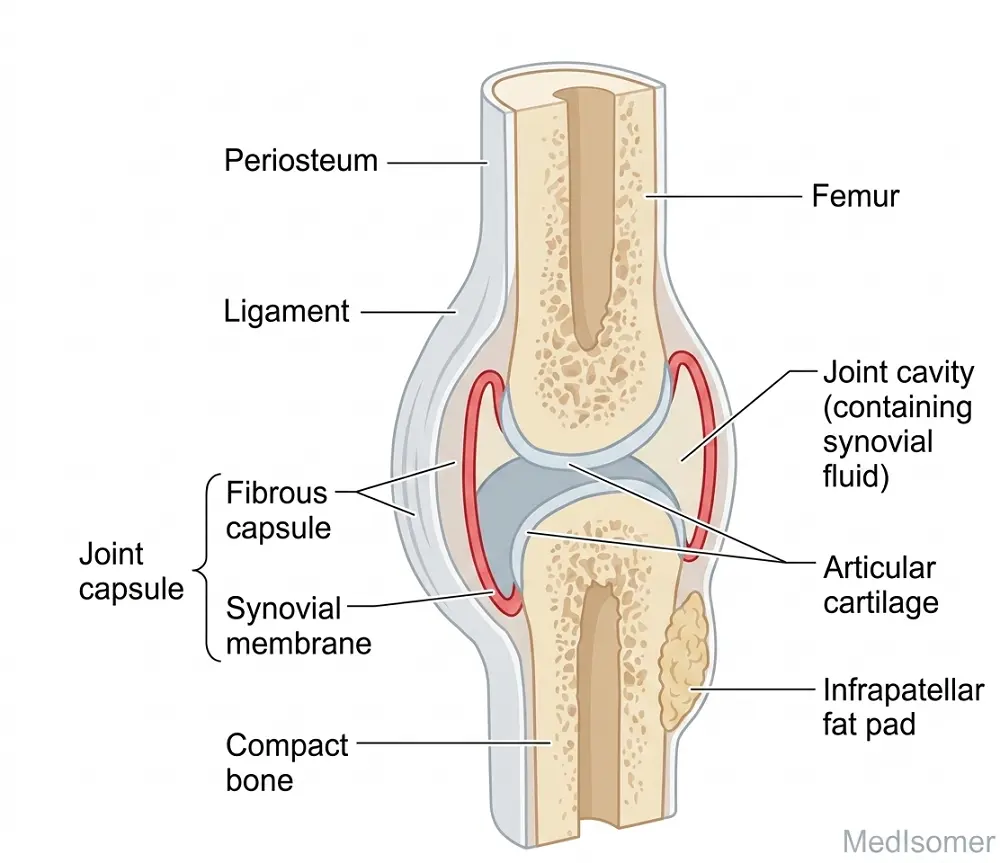
Synovial joints are further classified into six morphologic subtypes based on the shape of their articulating surfaces and the mechanical planes of movement allowed: • Plane (Arthrodial) Joints: Permit simple gliding movements across flat surfaces (e.g., acromioclavicular and intercarpal joints). • Hinge (Ginglymus) Joints: Uniaxial joints permitting flexion and extension alone (e.g., elbow/humeroulnar and knee joints). • Pivot (Trochoid) Joints: Uniaxial joints allowing rotation around a central axis (e.g., atlantoaxial and proximal radioulnar joints). • Condyloid (Ellipsoid) Joints: Biaxial joints permitting flexion, extension, abduction, adduction, and circumduction (e.g., metacarpophalangeal and radiocarpal joints). • Saddle (Sellar) Joints: Biaxial joints featuring concave and convex reciprocal surfaces, enabling precise multidirectional movement (e.g., 1st carpometacarpal joint of the thumb). • Ball-and-Socket (Spheroid) Joints: Multiaxial joints providing the highest degree of mobility through three perpendicular axes (e.g., glenohumeral and hip joints).
Vascular and Nerve Supply: Hilton's Law
Joints receive blood via rich periarticular anastomoses that circle the joint capsule to ensure unhindered blood flow during varied mechanical positions. The nerve supply of joints is governed by Hilton's Law, a critical clinical principle stating that the nerves supplying a joint also supply the muscles that move that joint and the skin covering their distal insertions. Joint capsules are heavily populated by proprioceptive sensory endings that monitor joint position and nociceptors that detect damaging mechanical stress or inflammation.
Summary
Summary of Core Concepts
To synthesize arthrology, recall that joints are fundamentally organized by structural composition (fibrous, cartilaginous, synovial) and functional mobility (synarthrosis, amphiarthrosis, diarthrosis). Synovial joints are uniquely defined by their fluid-filled joint cavities, articular cartilage, and dual-layered capsules. They are clinically sensitive structures where proper lubrication prevents mechanical breakdown. Understanding their mechanical classifications (uniaxial, biaxial, multiaxial) and neurological boundaries (Hilton's law) allows clinicians to trace and diagnose structural pathologies, sports injuries, and degenerative conditions like osteoarthritis effectively.